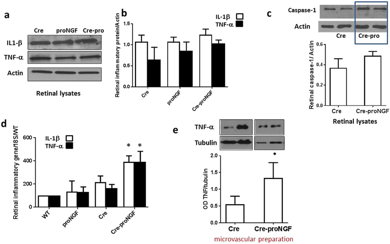
Fig. 5.
EC- proNGF overexpression induces expression of inflammatory markers. (a & b) Representative Western blots of TNFα, IL1β and actin in isolated retina. (d & e) Statistical analysis showed no significant change in cleaved-IL1β expression (17 kDa) or membrane-bound TNFα (26 kDa) expression among different animal groups (n = 5). (c) Representative image and statistical analysis of Western blots of retinal lysates showing no significant difference in cleaved caspase-1 (22 kDa) expression between Cre-proNGF and Cre-controls (n = 4). (d) Real-time PCR showed a significant increase in mRNA of IL1β (n = 5–6) and TNFα (n = 5–10) in retina isolated from Cre-proNGF mice compared to controls (*p < 0.05 vs WT, Cre and proNGF). (f) Representative of Western Blot and statistical analysis of membrane-bound TNF-α (26 kDa) expression in microvascular preparation showing significant (2-fold) increase in TNF-α from Cre-proNGF compared to Cre-controls(*p < 0.05 vs Cre, n = 4).

