Fig. 1.
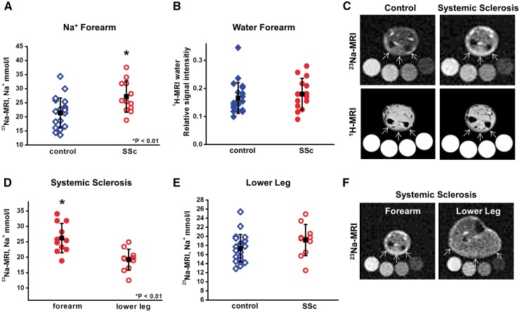
23Na-MRI detects increased Na+ in fibrotic SSc skin and discriminates between affected and non-affected skin
(A–C) Skin Na+ (A) and water content (B) of SSc patients (n = 12) and age-matched controls (n = 21) quantified by 23Na-MRI and 1H-MRI. (C) Representative23Na-MRI detects increased Na+ in fibrotic SSc skin and discriminates between affected and non-affected skin. 23Na- and 1H-magnetic resonance images of healthy (left) and fibrotic skin (right). Calibration tubes are placed below the forearm. Arrows indicate the site of measurement. (D) Skin Na+ content in fibrotic forearm vs non-involved lower leg in the same patients (n = 11). (E) Skin Na+ content of non-affected SSc skin vs healthy control patients (both lower leg). (F) Representative 23Na-magnetic resonance images of the forearm vs lower leg of an SSc patient.

