
Figure 2.
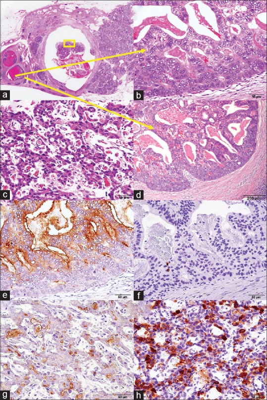
(a-d) Histological findings in the surgically resected specimen. (a) The main pancreatic duct in the head of the pancreas was occluded by a solid tumor. (b) A component in the branch duct was mainly a ductal component. (c) A component in the main pancreatic duct was mainly a neuroendocrine component (yellow square). (d) The transitional zone with minimal invasion into the pancreatic parenchyma was observed in the branch duct adjacent to the main pancreatic duct. (e-h) Immunohistochemically, a ductal component was positive for carcinoembryonic antigen (e), but not for synaptophysin (f), while a neuroendocrine component was positive for synaptophysin neuroendocrine component (g), not for carcinoembryonic antigen (h)