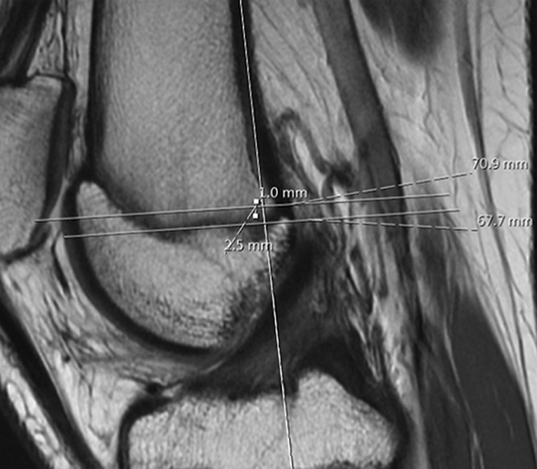
Figure 1.

Using the sagittal series on magnetic resonance imaging, a line is made in line with the posterior femoral cortex on the image with the best view of the anterior cruciate ligament. A second line is drawn at the most posterior aspect of the Blumensaat line perpendicular to the posterior cortical line. A third line is drawn at the most proximal aspect of the posterior femoral condyle that is perpendicular to the posterior cortical line. A point is measured that is 1 mm anterior to the posterior cortical line and 2.5 mm distal to the medial femoral condyle line, designated as the Schöttle point.
