Fig. 4.
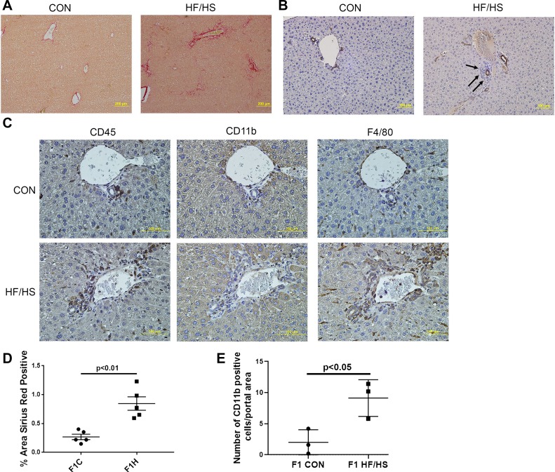
Periportal fibrosis and inflammation in offspring exposed to maternal high-fat/high-sucrose (HF/HS) diet. A: representative photomicrographs of picrosirius red (PSR) staining. B: representative photomicrographs of immunohistochemistry (IHC) for cytokeratin 19 (CK19). Arrows point to infiltrate around CK19-positive bile duct. C: representative photomicrographs of IHC for CD45, CD11b, and F4/80 in consecutive sections from maternal control (CON) and HF/HS offspring liver. D: quantitative analysis of PSR staining. E: no. of CD11b-positive cells per portal area (5 portal areas counted in each of 3 offspring/group). Quantitative data are presented as means ± SD with n = 5 (from 5 separate litters) in each group for PSR quantification and n = 3 (from 3 separate litters) in each group for CD11b cell counts. P values are as indicated.

