
Figure 4. Characterization of continuous polar growth during FtsZAT depletion.
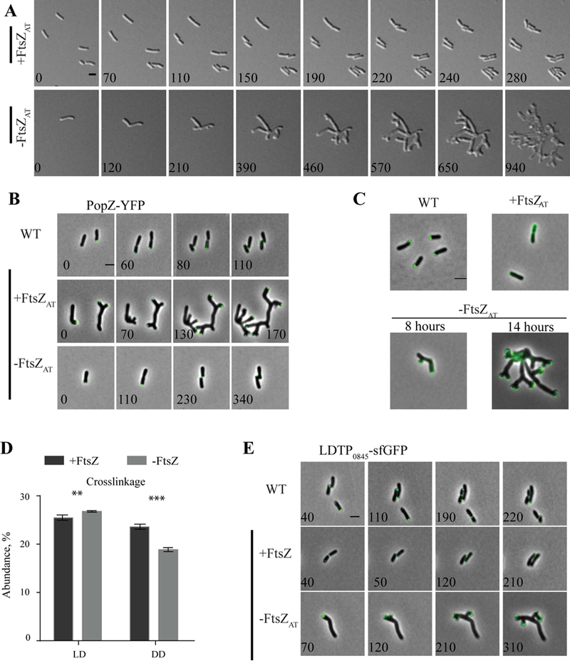
A) Timelapse microscopy of the ftsZAT depletion strain under inducing conditions (+FtsZAT, top panel) and depletion conditions (-FtsZAT, bottom panel). B) Timelapse microscopy showing PopZ-YFP localization WT, +FtsZAT and -FtsZAT. C) FDAA labeling of WT cells, and cells depleted of FtsZAT for 0 hours (+FtsZAT), 8 hours, and 14 hours. D) Abundance of total ld and dd crosslinkage in peptidoglycan isolated from ftsZAT depletion strain after induction (+FtsZAT, black bars) or depletion (-FtsZAT, grey bars) of FtsZAT for 14 hours. Data shown are the average abundance of each crosslinkage type and are taken from analysis of three independent biological samples. Statistical significance was calculated by t-tests and is indicated with an asterisk (P-value <0.05 (*), <0.005 (**), <0.001 (***)). E) Timelapse microscopy of LDTP0845-sfGFP in WT, +FtsZAT and -FtsZAT a. All scale bars are set to 2 µm.