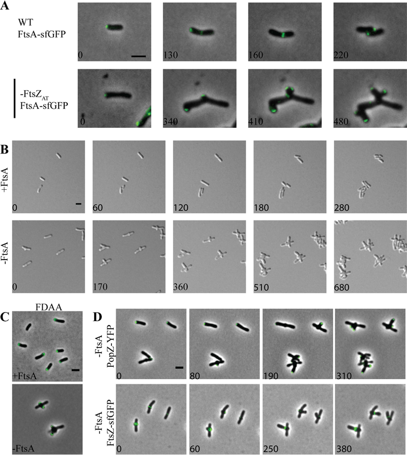
Figure 5. FtsA is not required for initiation of mid-cell growth.

A) FtsA-sfGFP localization in WT (top panel) and cells depleted of FtsZAT (bottom panel). B) Timelapse microscopy shows typical morphology when FtsA is induced and morphological changes in the absence of FtsA. C) FDAA labeling of the ftsA depletion strain after 8 hours of induction (+FtsA) or depletion (-FtsA). D) Timelapse microscopy of PopZ-YFP localization (top) and FtsZAT-GFP localization (bottom) during FtsA depletion. All scale bars are set to 2 µm.
