
Figure 1.
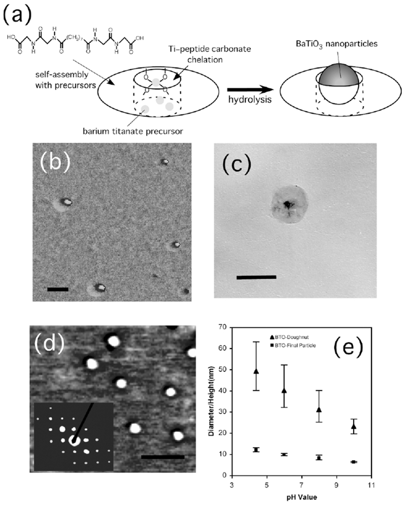
a) Illustration of the nanoring structure. Objects in this figure are not to scale and not all of the precursors that self-assemble with the peptide to form the nanoring are shown. b) Atomic force microscopy (AFM) phase image of barium titanate nanoparticles inside peptide nanoring templates. Scale bar = 50 nm. c) TEM image of a barium titanate nanoparticle inside the peptide nanoring template. Scale bar = 60 nm. d) AFM phase image of barium titanate nanoparticles after removal of peptide nanoring templates. Scale bar = 60 nm. Inset shows an electron diffraction pattern of the barium titanate nanoparticles with the (110), (111), (210), and (211) faces. e) Size distributions of peptide nanorings (▴) and barium titanate nanoparticles (■) as a function of pH of the growth solution.