
Figure 2.
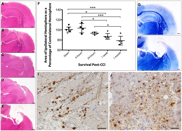
Progressive atrophy and associated axonal pathology post‐CCI in standard FFPE tissue. (A) H&E staining on sham animals reveals an absence of any focal pathology and a morphologically normal cortical surface. (B–E) H&E staining performed (B) 6 h, (C) 24 h, (D) 1 week and (E) 1 month after CCI injury reveals acute cortical contusion and intraparenchymal hemorrhage at the site of impact as well as progressive atrophy of the ipsilateral hemisphere. (F) Tissue atrophy quantified by measuring the area of the injured ipsilateral hemisphere as a percentage of the contralateral hemisphere. All individual data are presented in addition to the mean percentage area ± SD. In the sham group, gray data points indicate mice sacrificed 24 h after sham injury and black data points indicate mice sacrificed 1 month following sham injury, indicating an absence of change caused by craniectomy. (G–H) Luxol fast blue staining indicates loss of white matter and major cortical tissue loss at (H) 1 month following injury compared to (G) sham. (I–J) Swollen axonal profiles and varicosities immunroeactive for APP at (I) 24 h and (J) 1‐month post‐CCI. Scale bars: (A–E, G–H) 400 µm, (I–J) 25 µm. *P < 0.05, ***P < 0.001.