
Figure 3.
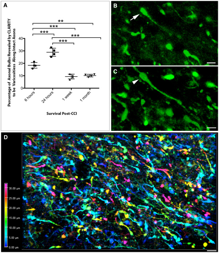
CLARITY reveals less axonal disconnection than previously thought following CCI. (A) Graph showing the percentage of APP+ swellings that appeared terminally disconnected when viewed in standard 8‐µm‐thick regions of tissue that were revealed to be connected varicose axonal swellings when all three dimensions were fully visible using CLARITY (**P ≤ 0.01, ***P ≤ 0.001). (B) Representative example of an APP+ apparent terminal axonal bulb (arrow) observed on an 8‐µm‐thick region of cleared tissue (maximum projection) at 24 h post‐CCI. (C) The same region of tissue as (B) re‐evaluated with all three dimensions now visible reveals that the apparent bulb is in fact a swelling that has two clear projections and thus represents a varicose swelling along an intact region of axon (arrowhead). (Alpha‐blended image) (D) Depth encoded three‐dimensional region of extensive APP+ axonal pathology in the peri‐contusional region 24 h post‐CCI. Extensive and large axonal swellings can be observed with complex morphologies comprising both disconnected axonal bulbs and varicosities along the intact length of axons. Scale bars: (B,C) 10 µm, (D) 50 µm.