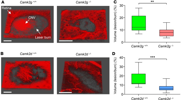
Figure 6. Laser-induced choroidal neovascularization (CNV) is reduced in CAMKIIγ- and -δ–KO mice.
CNV lesions were generated using laser photocoagulation and quantified by multiphoton imaging of isolectin B4–stained retinal-choroidal flatmounts. (A and B) Representative 3-D images of isolectin B4–stained CNV lesions from CAMKIIγ and -δ WT and homozygous KO mice. Large CNV lesions are clearly evident within laser burns of the WT mice. Scale bars: 10 µm. (C and D) Box-and-whisker plots (min, max, 25th–75th percentile, median) comparing CNV lesion volumes (expressed as a percentage of the total burn volume) between CAMKIIγ and -δ WT and KO mice. KO animals exhibited significantly less CNV than their WT counterparts. **P < 0.01 and ***P < 0.001 based on 2-tailed Student t test; n = 5–10 animals per group.

