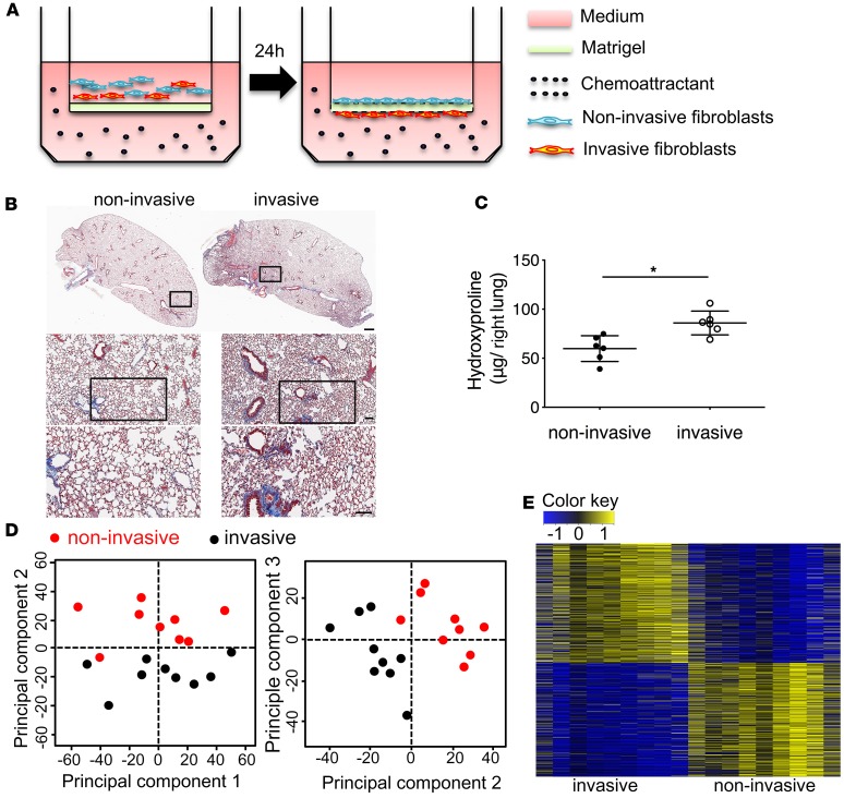
Figure 1. Invasive lung fibroblasts promoted interstitial lung fibrosis.
(A) Schematic representation of in vitro invasion assay. Lung fibroblasts were seeded in the upper part of transwells. Cells attached to the bottom of Matrigel-coated membrane after 24 hours were considered invasive fibroblasts. Cells remaining on top of the Matrigel-coated membrane were considered noninvasive fibroblasts. Invasive and noninvasive IPF lung fibroblasts (n = 9 per group) were isolated using the matrigel invasion assay. Masson’s trichrome staining of collagen on lung sections (B) and hydroxyproline content in lung tissues (C) from NSG mice 50 days after injection with invasive and noninvasive IPF lung fibroblasts on day 50 after fibroblast injection (n = 6 per group). Scale bars: 1 mm (top panel), 100 μm (middle and lower panels). (D) Principal component analysis of RNA-seq data. (E) Heatmap of all differentially expressed (DE) genes in RNA-seq data. A total of 1,405 DE genes were identified with FDR < 0.01 and |log2 FC| > 0.5; among them, 719 DE genes were upregulated, and 686 DE genes were downregulated. *P < 0.05 by Student’s t test (C).

