FIG 4.
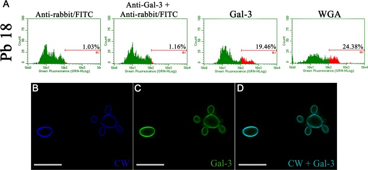
Gal-3 binds to P. brasiliensis cell wall. (A) P. brasiliensis Pb18 strain cultivated in YPD medium for 72 h at 37°C was resuspended in PBS and incubated sequentially with 40 μg/ml of Gal-3, an anti-Gal-3 antibody, and finally anti-rabbit IgG-FITC antibody. Binding was measured by flow cytometry; numbers inside histograms represent the percentages of positive cells recognized by Gal-3. WGA lectin-FITC (30 μg/ml) was the control for binding with cell wall (CW). (B) P. brasiliensis Pb18 strain cultured at 37°C for 72 h and incubated with Gal-3 was stained for observation of cell wall with calcofluor white (blue) (B) and Gal-3 with anti-Gal-3 antibody (green) (C). (D) Merged image (cell wall and Gal-3). The images represent a single section from a Z series stack. Scale bars, 10 μm. Data are representative of three experiments and a representative image is shown.

