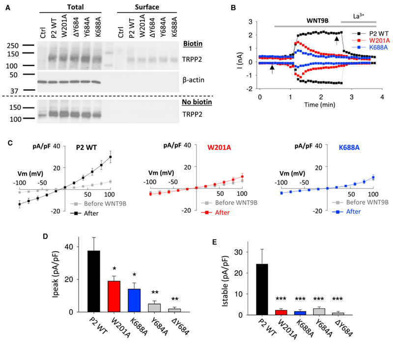
Figure 3. Roles of TRPP2 Residues W201 and K688 in WNT9B-Induced Whole-Cell Currents.

(A) Cell surface expression of WT TRPP2 and mutants W201A, ΛΥ684, Y684A, and K688A in HEK293 cells by transient co-transfection together with PKD1.
(B) Representative whole-cell current traces obtained at +100 and −100 mV before and after extracellular addition of WNT9B (0.5 μg/mL) or La3+ (100 μM) in CHO-K1 cells transiently co-expressing PKD1 with WT TRPP2, the W201A, or K688A mutant. The electrophysiological measurements were performed as described previously (Kim et al., 2016).
(C) Averaged steady-state I-V curves obtained before and ~2 min after application of WNT9B, at time points indicated by arrows in (B). WT TRPP2, n = 7 cells; W201A, n = 11 cells; K688A, n = 7 cells.
(D and E) Averaged peak (D) and steady-state (E) currents induced by WNT9B in CHO-K1 cells under the same experimental conditions as in (B). WT TRPP2, n = 7 cells; W201A, n = 11 cells; ΛY684, n = 8 cells; Y682A, n = 10 cells; K688A, n = 7 cells. *p < 0.05; **p < 0.01; ***p < 0.001.
Data are presented as mean ± SEM.
