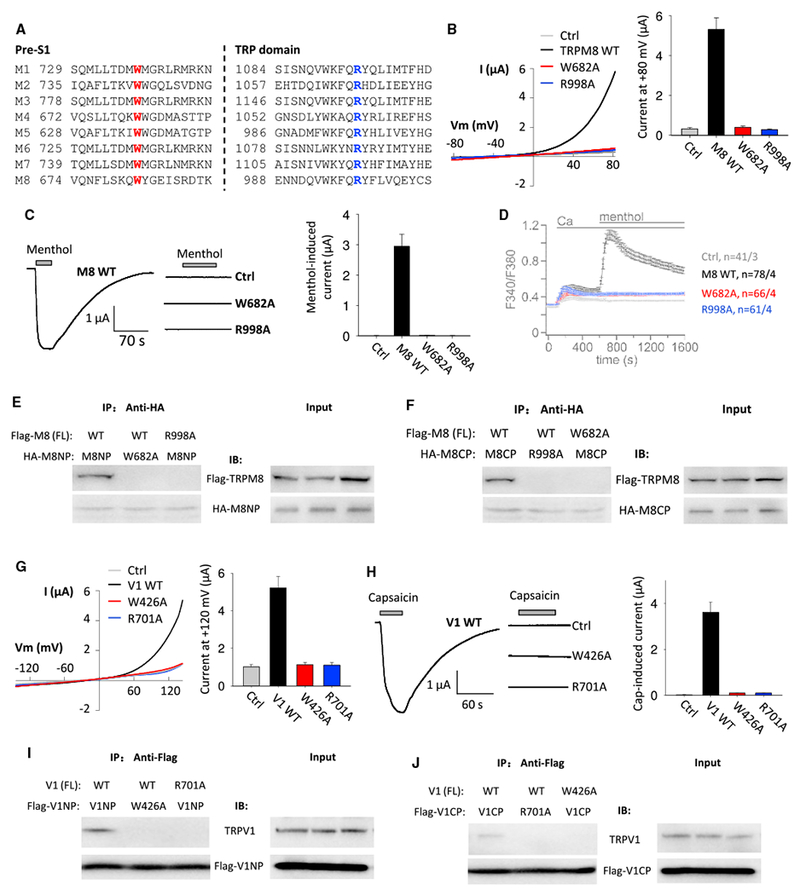
Figure 4. Roles of TRPM8 and TRPV1 Aromatic and Cationic Residues in the Pre-S1 and TRP-like Domains, Respectively, in the N-C Binding and Channel Function.

(A) Sequence alignment of human TRPMs pre-S1 and TRP-like domains.
(B) Left panel: representative whole-cell I-V curves obtained from oocytes expressing rat TRPM8 WT or a mutant channel in the presence of Na+-containing extracellular solution at RT. Ctrl, H2O-injected oocytes. Right panel: averaged currents at +80 mV from 12–18 oocytes of three batches are shown.
(C) Left panel: representative current traces obtained at −50 mV from oocytes expressing WT or a mutant TRPM8 before and after addition of 0.5 mM menthol. Right panel: averaged menthol-induced currents from 12–18 oocytes of three batches are shown.
(D) Ca2+-imaging measurements showing averaged fura-2 ratios obtained before and after Ca2+ (2 mM) and menthol (0.5 mM) addition to Na+-containing extracellular solution in HEK293 cells transiently co-expressing GFP with rat WT or a mutant TRPM8 or none (Ctrl) at 37°C.
(E and F) Representative co-IP data using oocytes expression, showing the interaction of Flag-tagged FL TRPM8 with HA-tagged TRPM8 N-terminal peptide (HA-M8NP; N642-K691; E) or C-terminal peptide (HA-M8CP; G980-F1029; F).
(G) Left panel: representative whole-cell I-V curves obtained from oocytes expressing rat WT or a mutant TRPV1 in the presence of the Na+-containing solution at RT. Ctrl, H2O-injected oocytes. Right panel: averaged currents at +120 mV from 10–16 oocytes of three batches are shown.
(H) Left panel: representative current traces obtained at —50 mV in rat WT or a mutant TRPV1 expressing oocytes before and after extracellular addition of capsaicin (15 μM). Right panel: averaged capsaicin-induced currents from 10–16 oocytes of three batches are shown.
(I and J) Representative co-IP data using oocytes expression, showing the interaction of rat FL TRPV1 with Flag-tagged TRPV1 N-terminal peptide (Flag-V1NP; D383-R432; I) or C-terminal peptide (Flag-V1CP; N687-D736; J).
Data are presented as mean ± SEM. See also Figures S3, S4, S5, and S6.
