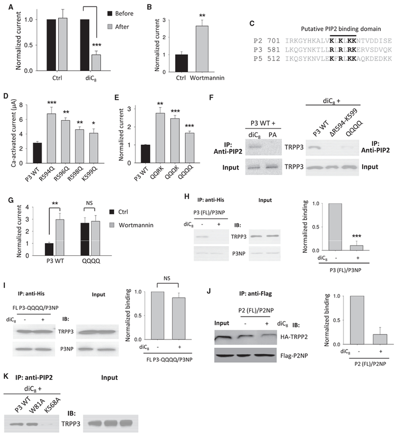
Figure 5. Inhibition of the TRPP3 Channel Function and N-C Interaction by PIP2.

(A) Averaged and normalized Ca-activated currents obtained from TRPP3-expressing oocytes before and after on-site injection of 25 nL water without (Ctrl) or containing diC8 (5 mM). Injection was performed with a third electrode after the initial current measurement. The second current measurement was performed 10 min after the injection. Currents were averaged from three independent experiments (with n = 12–15). ***p < 0.001.
(B) Averaged and normalized Ca-activated currents obtained from TRPP3-expressing oocytes pre-incubated with 10 μM wortmannin or DMSO (Ctrl) for 1 hr before measurements. **p < 0.01.
(C) Alignment of the C-terminal putative PIP2 binding domains in human TRPPs. Conserved cationic residues are highlighted.
(D) Averaged Ca-activated currents obtained from oocytes expressing WT or a mutant TRPP3 (n = 14–18). *p < 0.05; **p < 0.01; and ***p < 0.001.
(E) Averaged and normalized Ca-activated currents. QQRK, R594Q/R596Q double mutant; QQQK, R594Q/R596Q/R598Q triple mutant; QQQQ, R594Q/R596Q/ R598Q/K599Q quadruple mutant. **p < 0.01 and ***p < 0.001.
(F) Left panel: representative co-IP data showing interaction between diC8 PIP2 and TRPP3. Right panel: representative co-IP data show interaction of diC8 PIP2 with WT or a mutant TRPP3 expressed in oocytes. DR594-K599, TRPP3 deleted with fragment R594-K599. diC8 PIP2 or phosphatidic acid (PA, a negative control) was added to cell lysate to a final concentration of 15 μM. An anti-PIP2 antibody (sc-53412) from Santa Cruz Biotechnology was used for immuno-precipitation.
(G) Averaged and normalized Ca-activated currents obtained from oocytes expressing WT TRPP3 or the QQQQ mutant. Oocytes were treated with 10 μM wortmannin or DMSO (Ctrl) for 1 hr before measurements. **p < 0.01; NS, not significant.
(H) Left panel: representative co-IP data showing the effect of diC8 on the interaction of P3NP with FL TRPP3 in oocytes. diC8 was added in the cell lysis buffer to final concentration of 15 μM. Right panel: data from three independent experiments in left panel were quantified, averaged, and normalized. ***p < 0.001.
(I) Left panel: representative co-IP data showing the effect of diC8 on the interaction of P3NP with the TRPP3 QQQQ mutant. Right panel: data from three independent experiments in left panel were quantified, averaged, and normalized (NS, not significant).
(J) Left panel: representative co-IP data showing the effect of diC8 on the interaction of P2NP with FL TRPP2. Right panel: data from three independent experiments in left panel were quantified, averaged, and normalized.
(K) Representative co-IP data showing the interaction of PIP2 with expressed WT or a mutant TRPP3 in oocytes.
Data are presented as mean ± SEM. See also Figure S2.
