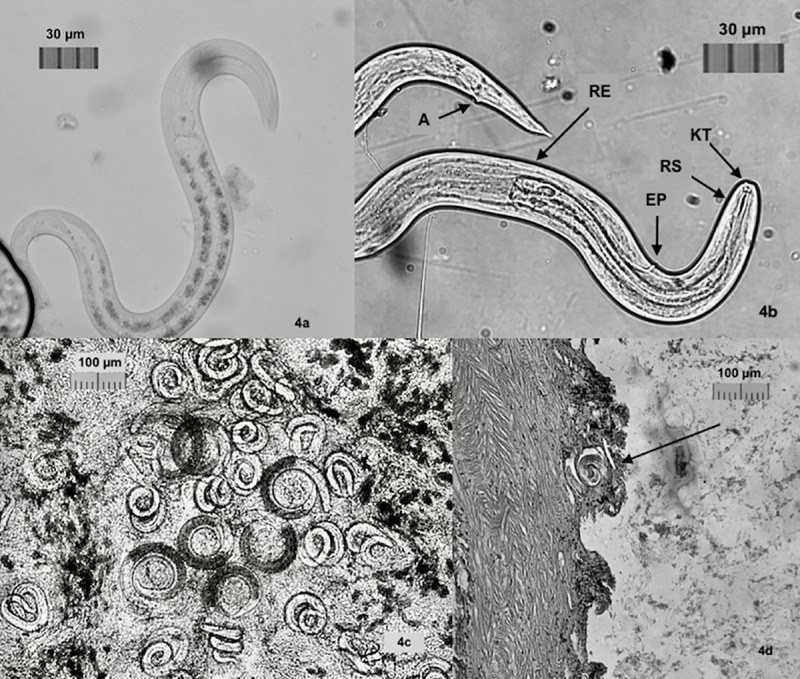
Fig 4. Images of A. cantonensis.

a. L1 larvae with distinctive junction of rhabditoid esophagus in the anterior section, posterior section intestinal cells dense with reflective granules (10μm). b. L3 larvae with knob-like tips (KT) and rod-like structure (RT) in head, clear division of rhabiditoid esophagus (RB), excretory pore (EP), and anus (A). Larvae were 28–35 μm in width to 450–490 μm in length. c. Tissue squash from P. martensi showing C-shaped L2 larvae with dark interiors. Larvae were 29–32 μm in width to 420–470 μm in length. d. Histology section of P. martensi showing coiled larvae very close to edge of the body wall.
