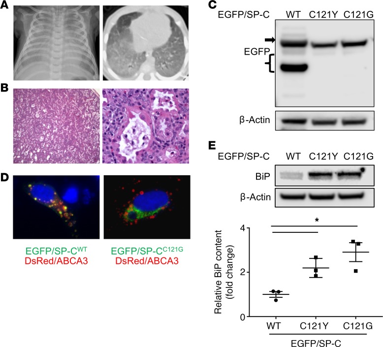
Figure 1. In vitro modeling of a clinical SFTPCC121Y mutation identifies mistrafficked pro-protein and epithelial ER stress with mutagenesis of codon 121 in SFTPC.
(A) Chest imaging of a 3-month-old patient with heterozygous SFTPCC121Y mutation shows bilateral diffuse hazy opacity on chest radiograph (left) and diffuse ground glass opacities on chest CT (right). (B) H&E staining of patient lung biopsy demonstrates diffuse abnormality in lobular architecture, with mild enlargement of airspaces and mild to moderate interstitial widening, airspaces filled by foamy macrophages, scattered neutrophils, and eosinophilic material suggestive of proteinosis (original magnification, ×4 left and ×20 right). (C) HEK293 cells were transiently transfected with either EGFP/SP-CWT or EGFP/SP-CC121 mutants as indicated, and cell lysates harvested at 48 hours were subjected to Western blotting for EGFP. An SP-C primary translation product (black arrow) was detected in all transfections; however, no processing intermediates (black bracket) were observed with either EGFP/SP-CC121 mutation. Representative of 3 independent experiments. (D) HEK293 cells transiently cotransfected with either EGFP/SP-CWT or EGFP/SP-CC121G and DsRed/ABCA3 and subjected to immunfluorescence microscopy, demonstrating colocalization of EGFP/SP-CWT with DsRed/ABCA3, but a reticular expression pattern of EGFP/SP-CC121G without DsRed/ABCA3 colocalization (original magnification, ×60). (E) Cell lysates of HEK293 cells 48 hours after transfection with either EGFP/SP-CWT or EGFP/SP-CC121 mutants as indicated were subjected to Western analysis for BiP (top). Representative Western blot of n = 3. Quantitative densitometry (bottom) normalized to β-actin showing increases in BiP with the cysteine mutants compared to EGFP/SP-CWT. n = 3. *P < 0.05 vs. EGFP/SP-CWT by 1-way ANOVA with Dunnett’s multiple comparison test.

