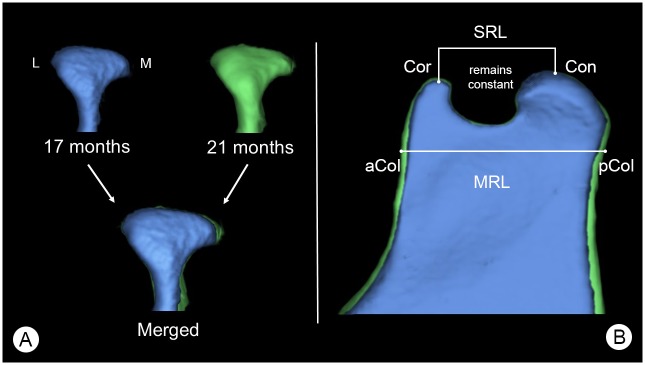
Fig 8. Growth changes of the mandibular condyle and superior ramus.
(A) Posterior view of a mandibular condyle of the same individual animal at 17 (blue) and 21 months (green) of age scaled to same size, showing changes in mandibular condyle volume (MCV) over time. Here: L = lateral aspect of the mandibular condyle and M = medial aspect of the mandibular condyle. (B) Lateral view of the superior area of the mandibular ramus, showing growth changes of the mandibular ramus. Here: Cor = coronion, Con = condylion, aCol = anterior point of the mandibular collum, pCol = posterior point of the mandibular collum. Parameters were: SRL = Cor-Con, MRL = aCol-pCol. Segmentations are presented at the same scale.

