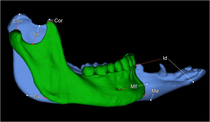
Fig 9. The different morphology of the minipig and human mandible.
3D renderings of an adult human mandible (green) and a mandible of a 21-months old Göttingen Minipig. Both segmentations are presented at the same scale. Where: Con = condylion, Cor = coronion, Sn = lowest point of the sigmoid notch, Go = gonion, Mf = posterior prominent mental foramen, Me = menton, Id = infradentale.

