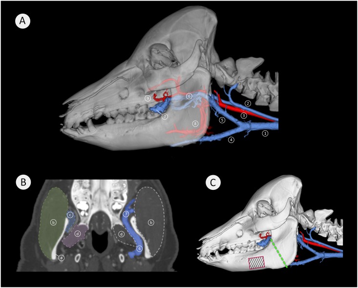
Fig 11. Vascular architecture medial to the mandibular ramus.
Image (A) is a lateral view of a semitransparent segmentation of a 21 months-old minipig head with associated major blood vessels of the neck and the mandibular region. Arteries are pictured in red and veins in blue. Here: (1) external jugular vein, (2) internal jugular vein, (3) common carotid artery, (4) linguofacial vein, (5) maxillary vein, (6) deep facial vein with maxillary artery, (7) deep facial vein traversing from medial to lateral, (8) lingual artery, (9) buccal artery. Image (B) shows a coronal view with the prominent deep facial vein (6) (in blue), adjacent to the medial aspect of the mandibular ramus (a). The vein has a diameter of approximately 6 mm and traverses from medial to lateral across the anterior aspect of the mandible (7). Here; (a) mandibular ramus, (b) masseter muscle, (c) temporal muscle insertion, (d) lateral pterygoid muscle, (6) deep facial vein, (7) deep facial vein traversing from medial to lateral. Image (C) is a lateral view of a 21 months-old minipig skull with associated large blood vessels of the neck and the mandibular region. Arteries are pictured in red and veins in blue. The green dashed line indicates the most common sectional plane used in experimental mandibular distraction osteogenesis procedures, the black-striped red rectangle indicates a common site for fixation plate placement in some experimental surgery (Fig 12B and 12C).

