Fig. 5.

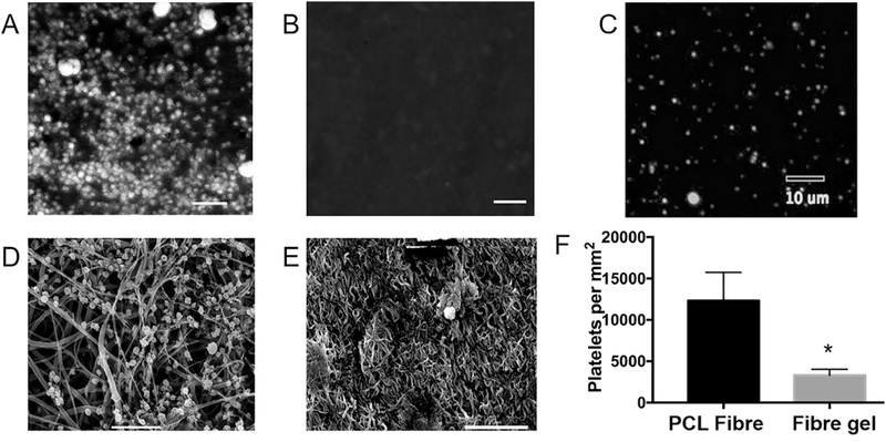
Platelet adhesion assay comparing two luminal graft surfaces. (A–C) Representative confocal images showing platelet adhesions on the PCL fibre surface (A) and coaxial PCL/gel fibre surface (B), as well as platelets in the testing solution (C). (D–E) Representative SEM images confirming that the coaxial PCL/gel surface (E) significantly decreased the adhesion and activation of platelets, when compared to the PCL surface (D). (F) Quantitative results from the platelet adhesion assay. All scale bars show 10 μm.
