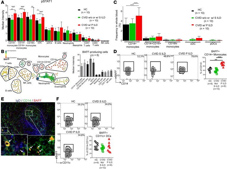
Figure 5. CD14+ monocytes are a prominent source of BAFF in CVID ILD.
(A) Whole blood was analyzed by mass cytometry, showing the highest phosphorylated STAT1 (p-STAT1) in monocytes and dendritic cells of CVID with progressive (P) ILD. CVID patients with no ILD and stable (S) ILD were grouped together because the cost of mass cytometry limited the number of samples that could be done and there was no difference in serum BAFF or STAT1 expression between these groups. (B) Monocyte and dendritic cells express the highest levels of intracellular BAFF in CVID ILD as detected by mass cytometry of 6 subjects. Displayed as density-normalized events (left) and plot of median intensity (right). (C) CD14+CD16– monocytes are elevated in CVID P ILD patients compared with CVID patients with stable (S) or no ILD and healthy controls (HCs), making CD14+ monocytes the most prevalent BAFF-producing subset. (D) A greater proportion of CD14+ monocytes produce BAFF in CVID P ILD compared with other CVID patients and controls. (E) Immunofluorescence of CVID ILD biopsies identified CD14+ monocytes as sources of BAFF in the lungs (white arrows). IgD+ B cell follicle highlighted with dotted white circle. Immunofluorescence shown is representative of 3 non-serial sections of lung biopsies from 3 CVID ILD patients. Original magnification, ×200 and ×1,600 (insets). (F) There was no difference in the proportion of lineage–CD11c+ dendritic cells producing BAFF between groups. *P < 0.05, **P < 0.01, ***P < 0.001, ****P < 0.0001. Error bars represent standard deviation. Multiple group comparison by Kruskal-Wallis test.

