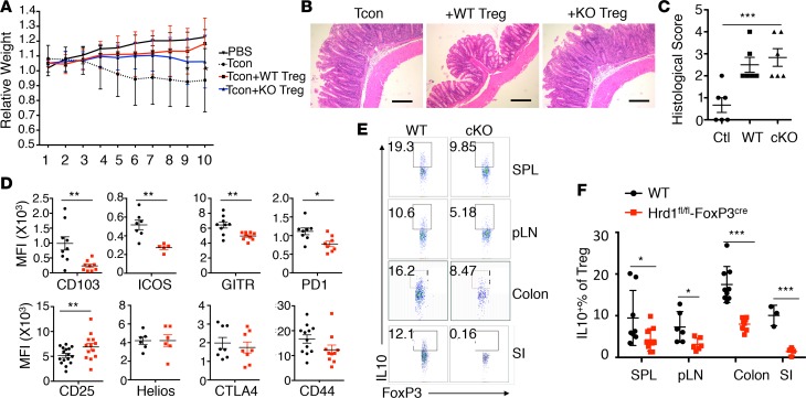
Figure 5. Reduced Treg suppressive function and altered Treg signature profiles Hrd1fl/fl-FoxP3cre mice.
(A) Colitis progression was assessed by body weight loss (n = 10 per group). (B and C) Representative images of the large intestine after H&E staining (B) and histological analysis (C) 10 weeks after adoptive transfer. Scale bars: 100 μm. Data are representative of 2 independent experiments with 5 mice per group in each experiment. (D) Expression of Treg-specific surface markers in thymic Tregs from WT and Hrd1fl/fl-FoxP3cre mice (n = 5–13 per group). (E and F) Representative images and quantification of IL-10–producing Tregs from WT and Hrd1fl/fl-FoxP3cre mice (n = 4–10 per group). Data are shown as mean ± SD. *P < 0.05; **P < 0.01; and ***P < 0.005 by 2-tailed Student’s t test.

