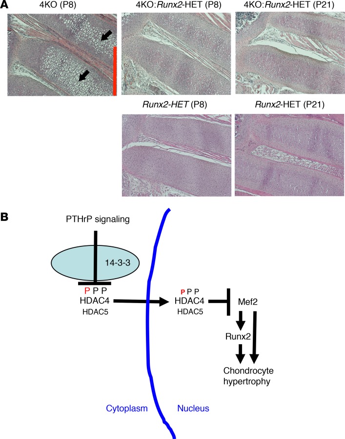
Figure 7. The Mef2/Runx2 signaling cascade is repressed by HDAC4/5 through PTHrP action.
(A) H&E staining of the anterior rib cartilage at P8 and P21 (original magnification, ×100). Mice at the same age are littermates. The Hdac4-KO mice die between P10 and P14. Black arrows indicate abnormal rib chondrocyte hypertrophy, which is cancelled by HET deletion of Runx2 (top images). Scale bar (red line): 500 μm. The Runx2-HET mouse exhibits normal rib phenotype (bottom images). (B) Proposed model for developmental regulation of chondrocyte hypertrophy. This model is derived from the data in this manuscript and those in previous reports (20, 21). PTHrP action increases HDAC4 nuclear translocation by decreasing phosphorylation of HDAC4 at the 14-3-3–binding sites. HDAC5 also mediates PTHrP signaling when HDAC4 expression is low. In the nucleus, HDAC4 and HDAC5 block the transcriptional activity of Mef2 through HDAC4/5’s Mef2-binding sites. Mef2 drives chondrocyte hypertrophy both by direct activation and through activation of Runx2 expression. Runx2 works downstream of HDAC4/Mef2 and HDAC5/Mef2 to drive chondrocyte hypertrophy.

