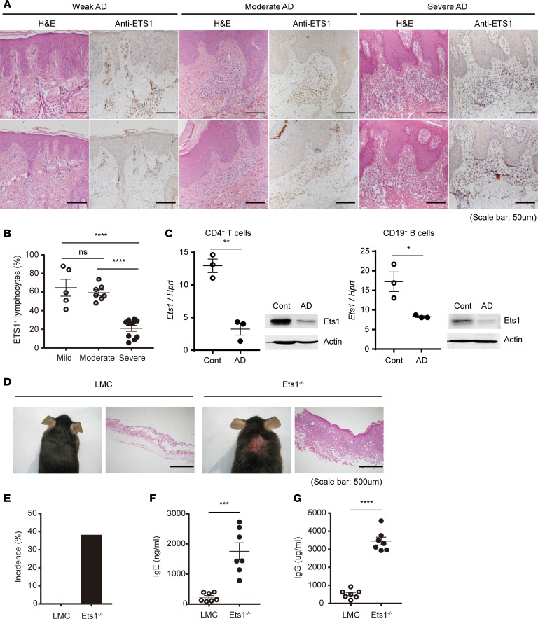
Figure 1. Ets1 expression is significantly reduced in skin lesion of severe AD patients and experimental animal model.
(A) H&E staining of the human skin biopsies confirmed the clinical diagnoses of weak, moderate, and severe atopic dermatitis (AD). Ets1+ lymphocytes were analyzed by IHC in the same tissues. (B) Frequency of Ets1+ lymphocytes from AD patient group with different disease severity is summarized in the graph. The data are expressed as mean ± SEM. ****P ≤ 0.0001; 1-way ANOVA. (C) After 4 weeks of AD induction in BALB/c mice, expression level of Ets1 was analyzed in peripheral CD4+ T and CD19+ B cells from skin-draining LNs by qPCR or immunoblotting. Quantification represents 3 independent experiments. (D) Representative phenotype of spontaneous AD-like skin inflammation developed in Ets1–/– mice (in C57BL/6 genetic background) under conventional conditions. (E) Incidence of AD-like skin inflammation among age-matched littermate controls (LMC) and Ets1–/– mice was calculated (n = 43 of LMC and 21 of Ets1–/– mice). (F and G) The total IgE and IgG levels in serum from mice groups at 12–16 weeks kept in conventional conditions were measured by ELISA (data comes from the analysis of 7 individual mice in each group). Error bars represent the mean ± SEM. *P ≤ 0.05; **P ≤ 0.005; ***P ≤ 0.0005; ****P ≤ 0.0001; Student’s t test.

