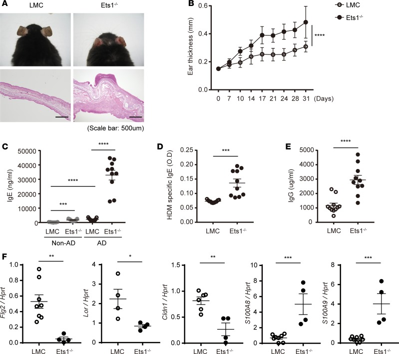
Figure 2. Ets1 deficiency promotes experimental AD pathogenesis under SPF conditions.
(A) Representative photographs of mouse ears from each group (upper) and H&E staining of the ear biopsies (lower) confirmed clinical symptoms of AD. (B) Ear thickness during the course of AD was measured 24 hours after DNCB or HDM extract application by using a dial thickness gauge. The data are expressed as mean ± SD. ****P ≤ 0.0001 (from day 17–31); 2-way ANOVA. (C–E) Total IgE (C), HDM allergen-specific IgE (D), and total IgG levels (E) in serum from the mouse groups were measured by ELISA. (F) RNA was collected from the cells of ear tissues, and the relative levels of filaggrin, claudin, loricrin, S100A8, and S100A9 were evaluated by qPCR after normalization with Hprt level. Data represent results from 3 independent experiments. Error bars represent the mean ± SEM. *P ≤ 0.05; **P ≤ 0.005; ***P ≤ 0.0005; ****P ≤ 0.0001; Student’s t test.

