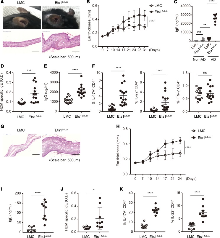
Figure 3. Specific deletion of Ets1 in mature T cells (Ets1ΔdLck) promotes AD pathogenesis by enhancing Th17 and Th2 responses.
(A) Representative photographs of mouse ears from each group (upper) and H&E staining of the ear biopsies (lower) confirmed the clinical symptoms of AD. (B) Ear thickness during the course of AD was measured. The data are expressed as mean ± SD. ****P ≤ 0.0001 (from day 17–31); 2-way ANOVA. (C–E) Total IgE (C), HDM allergen-specific IgE (D), and total IgG levels (E) in serum from the mouse groups were measured by ELISA. (F) Ex vivo isolated total lymphocytes from skin-draining LNs under AD were given PMA and ionomycin stimulation with GolgiStop or GolgiPlug for 4 hours. Intracellular staining of IL-17, IL-22, and IFN-γ by CD4-gated T cells were analyzed. (G) Naive CD4+ T cells were isolated from LMC and Ets1ΔdLck mice by means of fluorescence-activated cell sorting and transferred i.v. into Rag1−/− mice (5 × 10⁵/mouse) with WT CD45.1+CD19+ B cells (2 × 10⁶/mouse). AD was induced the day after cell transfer. H&E staining of the ear biopsies confirmed clinical symptoms of AD. (H) Ear thickness during the course of AD was measured. The data are expressed as mean ± SD. **P ≤ 0.005; ***P ≤ 0.0005; ****P ≤ 0.0001 (from day 17–24); 2-way ANOVA. (I and J) The total IgE (J) and HDM allergen-specific IgE levels (J) in serum from the mouse groups were measured by ELISA. (K) Intracellular staining of IL-17 and IL-22 by CD4-gated T cells were analyzed. Data represent results from 3–4 independent experiments. Error bars represent the mean ± SEM. *P ≤ 0.05; ****P ≤ 0.0001; Student’s t test.

