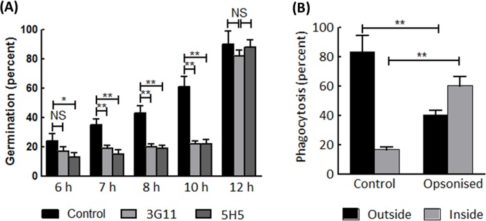
Fig 6. Antifungal activity of mAbs 3G11 and 5H5.
(A) A. fumigatus germination inhibition assay. Conidia were treated with 3G11 or 5H5 (PBS buffer was used as a control) and placed on Sabouraud agar medium spread on a glass slide. Conidial germination was monitored after 6 h of incubation at 37°C. The incubation time is shown on the X-axis. The assay was repeated three times, and at least hundred conidia were counted each time. (B) Phagocytotic assay. Swollen conidia (FITC-labelled) were treated with 3G11 for 60 min followed by feeding to human monocyte derived macrophages (HMDM). After a further 60 min of incubation at 37°C in a CO2 incubator, non-phagocytosed conidia were differentiated from phagocytosed FITC-labelled conidia by calcofluor white staining. Images were taken from different places to count at least one hundred swollen conidia in each experiment. Statistical analysis was performed by one-way ANOVA; *, p<0,05 and **, p<0,005.

