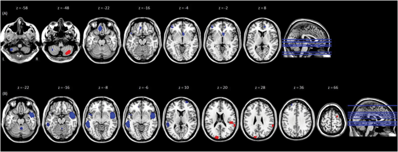
Fig. 2.
Results of the meta-analysis of (A) DMN to whole-brain functional connectivity, and (B) SN to whole-brain functional connectivity, in FEP individuals, relative to controls. Brain regions that showed significant differences in functional connectivity with (A) the DMN seed network, and (B) the SN seed network, in individuals with first-episode psychosis (FEP), relative to healthy controls (HC). MNI z coordinates are displayed at the top of the figure. Peaks appear from left to right in the (A) left cerebellum (P = .0026); right inferior semi-lunar cerebellar lobule (P = .00033); medial orbital gyrus (P = .00048); left superior temporal gyrus (P = .0026); ventral anterior cingulate gyrus (P = .00077); the inferior frontal gyrus (P = .0027); and the dorsal anterior cingulate gyrus (P = .0018); (B) cerebellum/culmen (P = .0033); right middle temporal gyrus (P = .000015); left planum polare (P = .0029); left middle temporal gyrus (P = .000034); middle frontal gyrus (P = .0011); superior temporal gyrus (P = .00038) and posterior transverse temporal gyrus (P = .00068); occipital gyrus (P = .0004); left superior frontal gyrus (P = .0027); right superior frontal gyrus (P = .00054). For color, please see the figure online.

