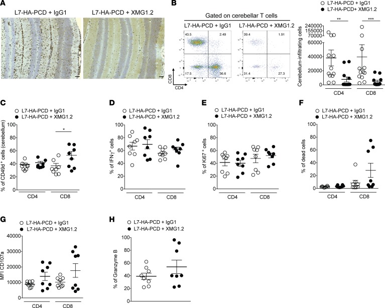
Figure 4. Cerebellar infiltration by T cells is reduced in PCD mice treated with anti–IFN-γ mAb.
(A) Representative IHC for CD3+ cells (brown) with nuclear counterstaining with hematoxylin (blue) on cerebellar sections from L7-HA-PCD mice treated with XMG1.2 (neutralizing anti–IFN-γ mAb) or control IgG1. Scale bar: 100 μm. (B) Left: representative flow cytometry staining at day 16 for CD4 and CD8 gated on live cerebellum-infiltrating T cells (CD90.2+) from L7-HA-PCD mice treated with XMG1.2 or IgG1. Right: absolute numbers of CD4 and CD8 T cells infiltrating the cerebellum of L7-HA-PCD mice treated or not with XMG1.2 (n = 12 per group, 3 independent experiments; Mann-Whitney U test, **P < 0.01; ***P < 0.001). (C) Expression of the α4 integrin (CD49d) on CD4 and CD8 T cells from the cerebellum of L7-HA-PCD mice treated with XMG1.2 or IgG1 (n = 8 per group, 2 independent experiments; Mann-Whitney U test, *P < 0.05). Frequency of IFN-γ+ cells (D), proliferating (Ki67+) cells (E), and dead cells (F) among cerebellum-infiltrating CD4 and CD8 T cells of L7-HA-PCD mice treated with IgG1 or XMG1.2 (n = 8 per group, 2 independent experiments). Geomean of CD107a cells (G) and frequency of Granzyme B+CD8+ cells (H) among cerebellum-infiltrating T cells of L7-HA-PCD mice treated with IgG1 or XMG1.2 (n = 8 per group, 2 independent experiments).

