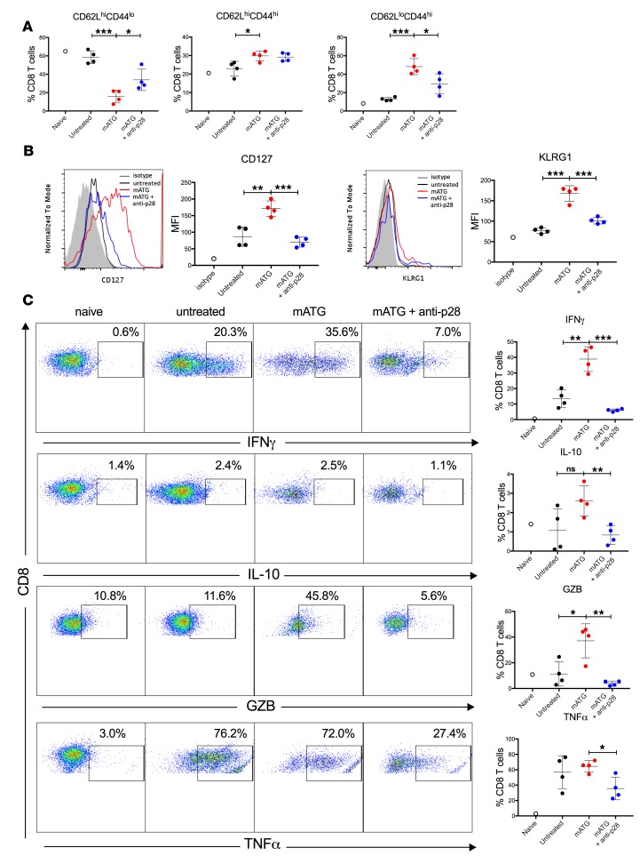
Figure 7. IL-27 neutralization alters the phenotype of reconstituted CD8+ T cells.
C57BL/6J mice were transplanted with BALB/c heart allografts and either left untreated or treated with either mATG (1 mg i.p. on days 0 and 4) plus anti-p28 mAb (clone MM27-7B1, 0.25 mg i.p. on days 6 and 11 after transplant) or mATG alone. Recipients were sacrificed on day 12 after transplant and splenic T cells were evaluated by flow cytometry. (A) Percentages of CD8+ T cells with naive (CD62LhiCD44lo), Tcm (CD62LhiCD44hi), or Tem (CD62LloCD44hi) phenotype. (B) Expression of CD127 (IL-7Rα, left) and KLRG1 (right) after gating on CD8+ cells. The results are shown as representative histograms (shaded, isotype control staining; black, untreated; red, mATG; blue, mATG + anti-p28) and MFI values. (C) Intracellular staining for IFN-γ, IL-10, Granzyme B (GZB), and TNF-α after gating on CD8+ cells. The results are shown as representative dot plots (left) and percentages of CD8+ T cells expressing respective cytokines (right). n = 4 mice per group, error bars represent SD. *P < 0.05, **P < 0.01, ***P < 0.001; ns, P ≥ 0.05 by multiple t tests.

