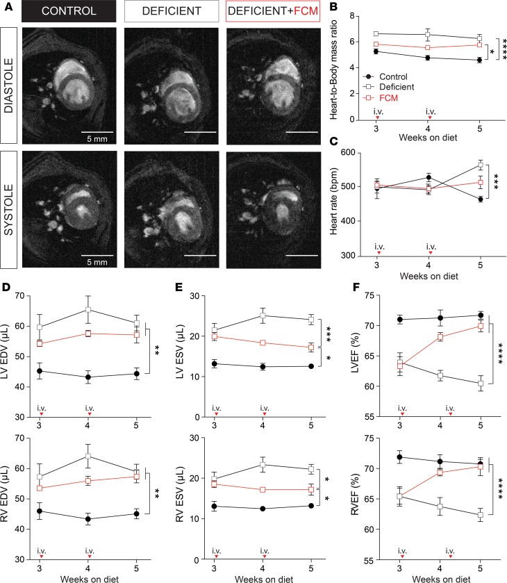
Figure 2. Effects of iron-deficiency anemia on cardiac function.
(A) Representative Cine-MR images obtained from control, deficient, and iron supplemented animals scanned after 5 weeks of diet, showing 1 slice (out of 11 collected in the z axis) at systole and diastole of 1 exemplar cardiac cycle. (B) Heart/body mass ratio and (C) heart rate. Left and right ventricular (D) end-diastolic volume (EDV), (E) end-systolic volume (ESV), and (F) ejection fraction (LVEF, RVEF) calculated from volume-rendering of MR images (n = 6 animals/group). Arrows indicate point at which FCM was injected. *P < 0.05, **P < 0.01, ***P < 0.001, ****P < 0.0001. P values were determined using unpaired Student’s t test (2-tailed).

