
Fig. 5.
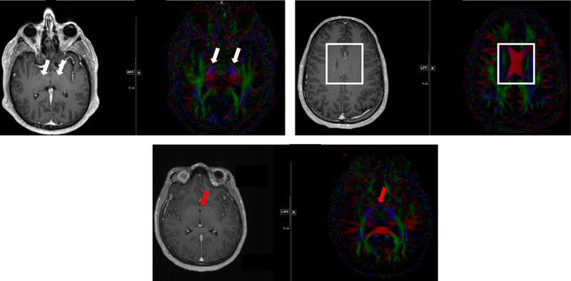
Representative MRIs and diffusion tensor imaging (DTI) showing white matter tracts for consideration in glioblastoma. Red indicates left-right pathways, blue indicates head-foot directionality, and green highlights anterior-posterior. In (A) the white arrows highlight blue ascending/descending white matter tracts at the level of the thalamus. These tracts are often not respected caudally, with inappropriate sparing of at-risk tissue below the level of the tentorial incisure. (B) The body of the corpus callosum is highlighted as indicated by the red pathways crossing midline. The falx cerebri is often inaccurately assumed to extend caudally to the top of the lateral ventricles. (C) Red arrows highlight the anterior commissure, with the DTI image demonstrating fibers in red that represent a white matter tract crossing midline. Similar fibers exist in the posterior commissure and variably in the interthalamic adhesion.