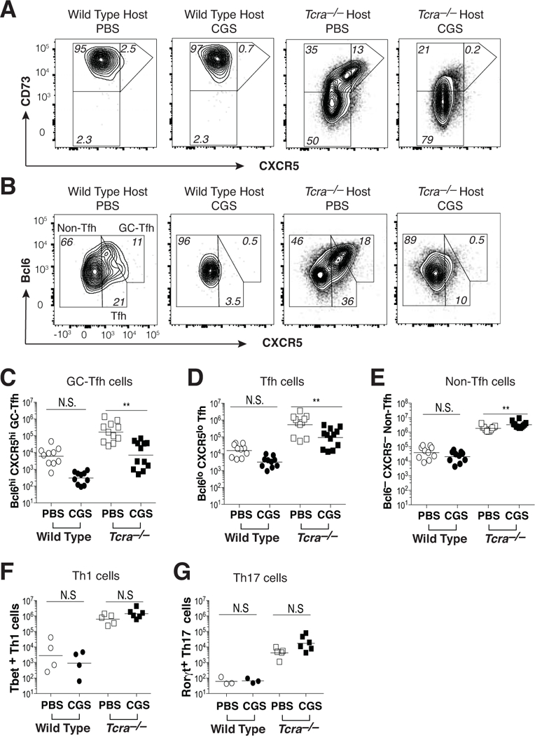
Figure 4. A2aR agonist treatment blocks Tfh and GC-Tfh cell differentiation during self-antigen recognition.

KRN CD4 T cells were transferred into wild type and Tcra–/– F1 hosts, and then mice were treated twice daily for 10 days with either CGS or PBS. (A) Expression of CD73 and CXCR5 on conventional Foxp3– KRN T cells. (B) Bcl6 and CXCR5 expression by conventional Foxp3– KRN T cells. (C-E) Aggregate numbers of Bcl6hi CXCR5hi (GC-Tfh) (C), Bcl6lo CXCR5lo (Tfh) (D), and Bcl6– CXCR5– (non-Tfh) (E) conventional Foxp3– KRN T cells. (F, G) Th1 (Tbet+) (F) and Th17 (RORγt+) (G) lineage T cells within the non-Tfh (Bcl6– CXCR5–) fraction of conventional Foxp3– KRN T cells. Data are representative of 3–4 independent experiments (n = 4–12 mice). **P < 0.01 by the Students t-test. N.S. = not significant.
