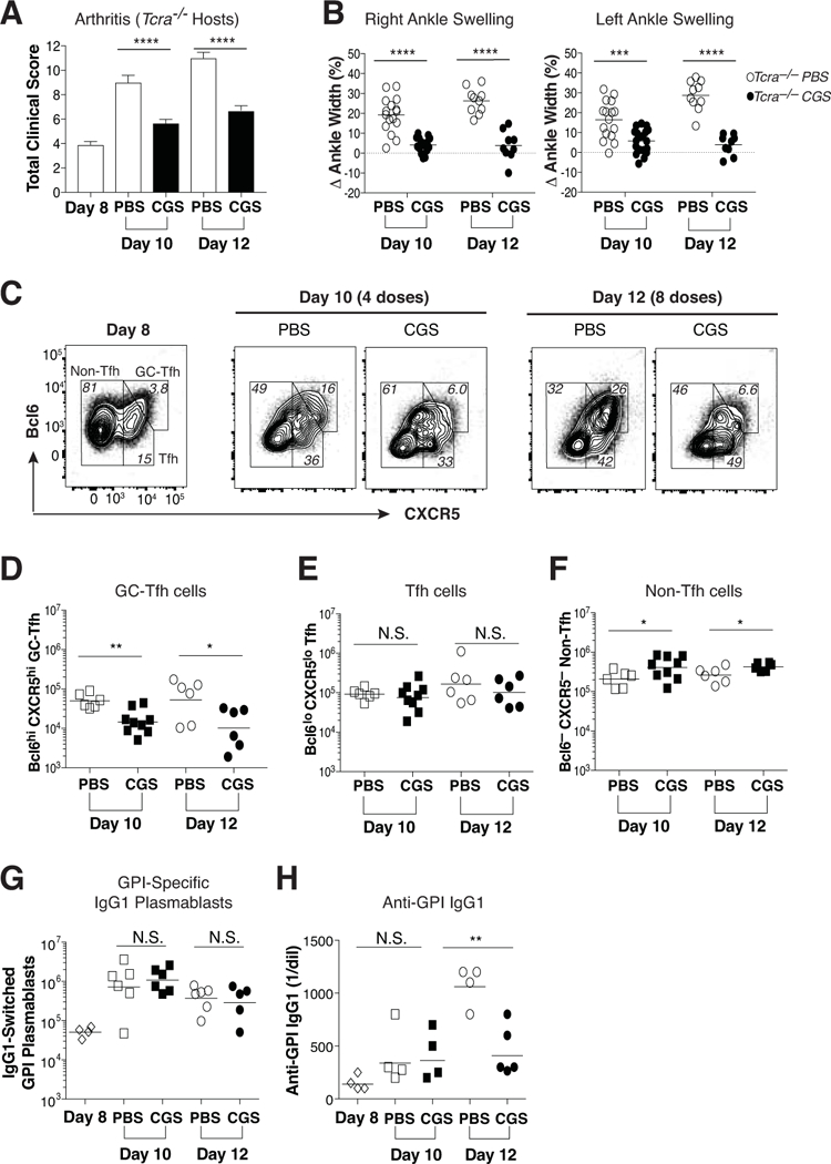
Figure 6. Treatment with CGS arrests GC-Tfh differentiation and blocks the progression of autoimmune arthritis.

Wild type KRN CD4 T cells were adoptively transferred into Tcra–/– F1 hosts. Beginning on day 8, arthritic mice were injected twice daily with CGS or PBS alone. (A) Mean total clinical disease activity scores (±SEM) observed on days 8, 10, and 12. (B) Percent change in right and left ankle swelling/size from the day 8 measurements. Data are representative of at least three independent experiments (n = 9–16). ***P < 0.001 by Mann Whitney U test and Students t-test, respectively. (C) Representative Bcl6 and CXCR5 expression patterns for conventional Foxp3– KRN T cells at days 8, 10, and 12. (D-F) Absolute numbers of GC-Tfh (Bcl6hi CXCR5hi) (D), Tfh (Bcl6lo CXCR5lo) (E), and non-Tfh (Bcl6– CXCR5–) (F) phenotypes among the conventional Foxp3– KRN T cells observed on days 10 and 12. (G, H) Host polyclonal B220dim intracellular Ig (H+L)hi GL7– IgG1+ GPI-specific plasmablasts (G) and serum anti-GPI IgG1 antibody titers (H) measured at days 8, 10, and 12. These data are representative of at least three independent experiments (n = 4–9). *P < 0.05 and **P < 0.01 by Students t-test. N.S. = not significant.
