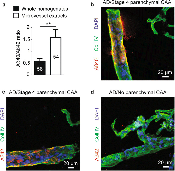
Fig. 2.
Localization of Aβ peptides in microvessel extracts from the parietal cortex. a) Concentrations of Aβ40, Aβ42 and Aβ40/Aβ42 ratios were determined in brain microvascular extracts by ELISA. A 3-fold higher Aβ40/Aβ42 ratio was observed in brain microvessel extracts compared to whole homogenates from the same parietal cortex samples. Data are represented as mean ± S.E.M. Sample size is indicated in the graph bars. Statistical analysis: unpaired Student’s t-test. ** p < 0.01. b-d) Immunolabeling of Aβ40 and Aβ42 following formic acid pretreatment revealed that both peptides accumulated on larger vessels at stage 4 parenchymal CAA in parietal cortex while no immunoreactivity was observed at stage 0 parenchymal CAA. Markedly, for both stages, no signal was found in smaller capillary-like vessels. Magnification used: 20X. Scale bar: 20 μm

