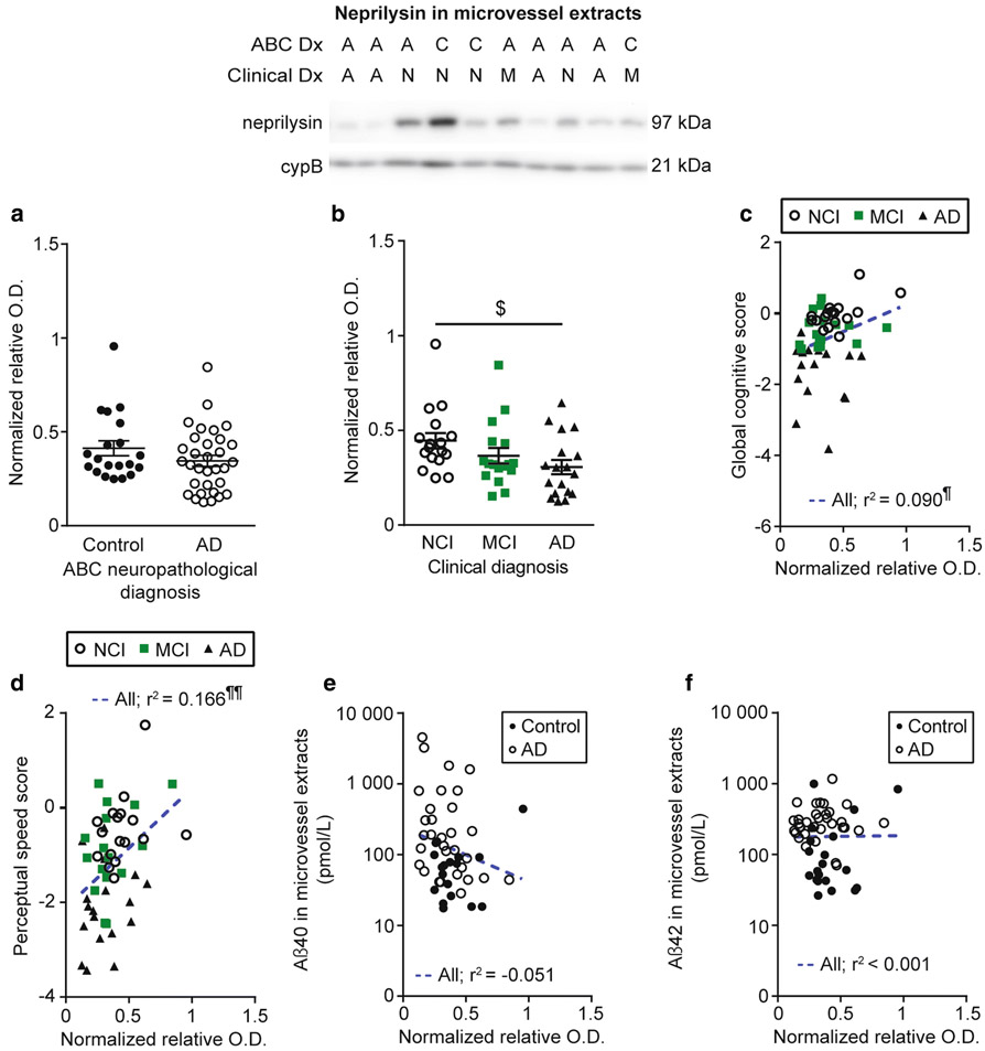
Fig. 6.
Neprilysin levels are reduced in brain microvessels from individuals with AD and correlated to cognitive function and Aβ40. Neprilysin levels in microvessel extracts were determined by Western blot. Data were normalized with cyclophilin B. No difference was found when participants were divided according to their AD neuropathological assessment (panel a) while a significant decrease was observed in individuals with AD based on clinical diagnosis (panel b). All samples, loaded in a random order, were run on the same immunoblot experiment. Consecutive bands are shown. Data are represented as scatterplots. Horizontal bars indicate mean ± S.E.M. Statistical analysis: Kruskal-Wallis ANOVA; $ p < 0.05. Neprilysin levels in microvessel extracts were positively associated to global cognition and perceptual speed (panels c and d). A trend towards a negative correlation between vascular neprilysin levels and Aβ40 (panel e) was noted while no significant association was found with Aβ42 (panel f). Statistical analysis: Pearson correlation coefficient. ¶ p < 0.05, ¶¶ p < 0.01. Abbreviations: A/AD, Alzheimer’s disease; ABC Dx, ABC neuropathological diagnosis; C, control; Clinical Dx, clinical diagnosis; cypB, cyclophilin B; M/MCI, mild cognitive impairment; N/NCI, healthy controls with no cognitive impairment; relative O.D., relative optical density

