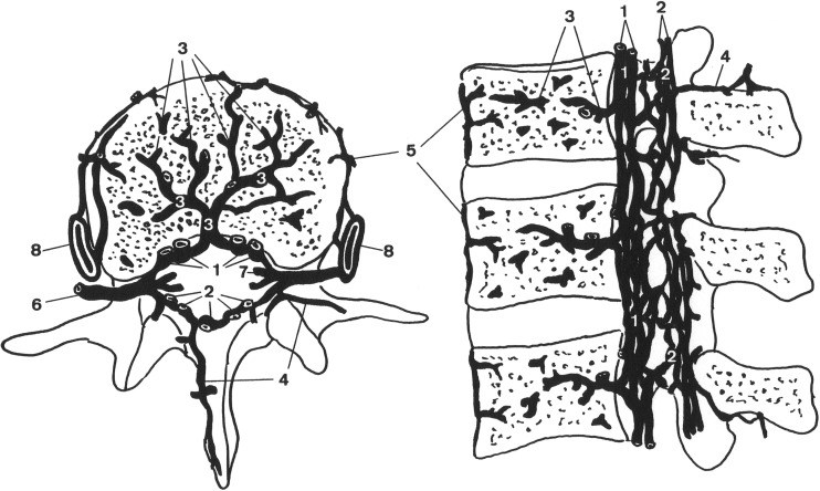
Fig. 3.
Schematic representation of the vertebral venous system (VVS) at the lumbar area showing the anterior internal vertebral venous plexus (1), posterior internal vertebral venous plexus (2), basivertebral veins (3), posterior external vertebral venous plexus (4), anterior external vertebral venous plexus (5), intervertebral vein (6), radicular vein (7), and the ascending lumbar vein (8). Reproduced from Groen et al. [62] with permission from Lippincott Williams & Wilkins

