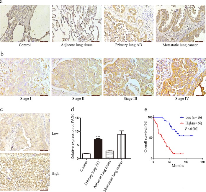
Fig. 1. PAX6 expression is correlated with poor prognosis in lung cancer.
a, d Representative images of IHC staining for PAX6 in samples from human NSCLC tissue arrays (magnification, 400×). Semi-quantitative results of PAX6 expression levels in lung cancer tissue arrays. ***P < 0.001, two-tailed Student’s t-test. b, c Representative images of PAX6 IHC staining in samples from a human NSCLC tissue array (magnification, 400×). e Kaplan–Meier analysis of overall survival of 92 NSCLC patients. Each subgroup was divided into low- (below or equal to the median value) and high-PAX6 expression groups (above the median value). P < 0.0001, log-rank test

