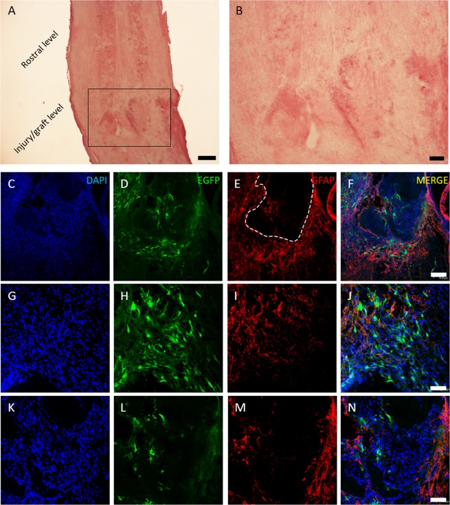
Figure 8.
MSC and CS/β-GP hydrogel transplantation. Hematoxylin/eosin staining shows the uninjured rostral spinal level and the lesion/graft site (A); in (B) a higher magnification of the injury level. In blue DAPI nuclei (C-G-K), in green EGFP-positive MSCs (D-H-L), in red astrocytes forming the glial scar at the lesion site (E-I-M) and the three previous images overlapped (F-J-N). In (C–F) the lesion area at low magnification shows astrocyte distributed around the glial cyst (dashed line in E). At higher magnification MSCs are visible around (H) and inside the lesion (L). Scale bar: (A) 300 μm, (B) 100 μm, (C–F) 70 um, (G–N) 40 um.

