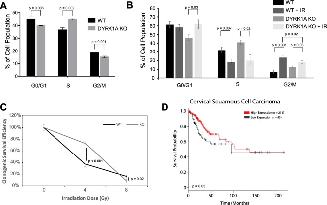
Figure 5.
Knockout of DYRK1A promotes radioresistance and cell survival. (A) Quantification of proliferating HeLa cells by cell cycle phase: flow cytometry analysis of propidium iodide staining and BrdU incorporation; 10 µM BrdU was pulsed 60 minutes prior to harvest. n = 3 (student’s t-test). (B) Quantification of HeLa cells by cell cycle phase 18 hours following 4 Gy of IR: flow cytometry analysis was done using propidium iodide staining of either WT or DYRK1A KO HeLa cells. n = 3 (student’s t-test). (C) Results of clonogenic survival assay show increased radioresistance of DYRK1A KO cells above WT survival following 4 Gy of IR. Cell colonies were counted using clono-counter java package 12 days following initial radiation. (D) Kaplan-Meier survival analysis of cervical squamous cell carcinoma tumors generated through KM plotter74,75. Patients with high DYRK1A expression in their tumors had increased survival probability over those with low DYRK1A expression.

