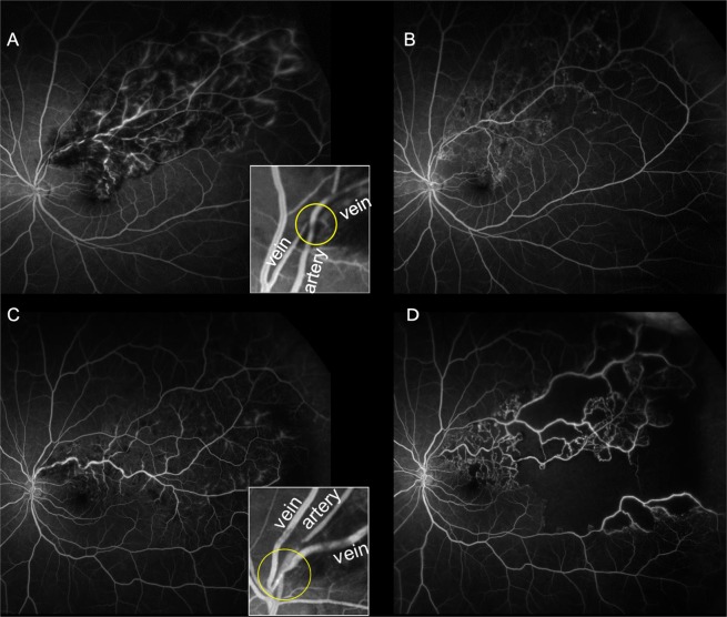
Figure 2.
Enlargement of retinal nonperfusion areas (NPAs) according to the anatomical position of the retinal vessels at affected arteriovenous crossings in eyes with branch retinal vein occlusion (BRVO). Ultrawide-field fluorescein angiography showing an arterial overcrossing pattern (A,B) and a venous overcrossing pattern (C,D). (A,C) Images at baseline. (B,D) Images at month 12. Images inset in panels A, and C show enlargements of the affected crossing sites. In images for a representative case of venous overcrossing (C,D), a marked increase in NPA, especially in the periphery, can be observed. In contrast, NPA enlargement is unremarkable in representative images for a case of arterial overcrossing (A,B).

