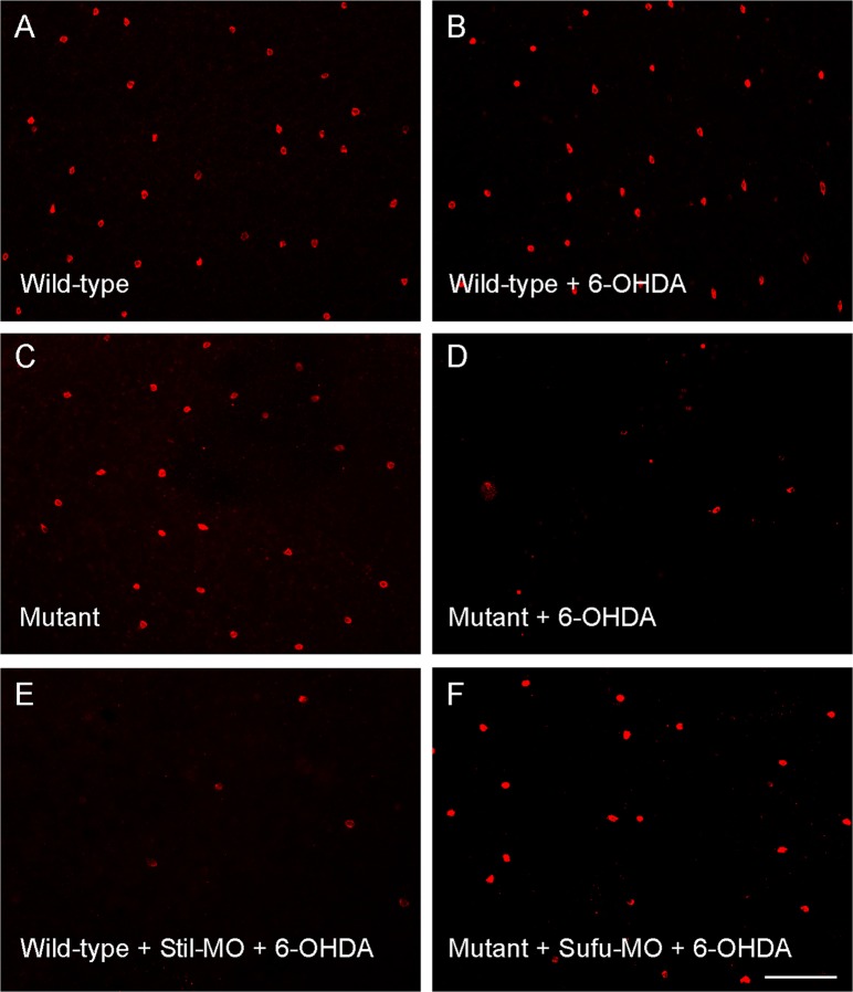
Fig. 2. Fluorescent images of flat-mounted zebrafish retinas that show the DA cells (labeled with antibodies against tyrosine hydroxylase) after treatment with sub-toxic 6-OHDA.
a, b Wild-type retinas that received sham or sub-toxic 6-OHDA treatment. No differences in the number of DA cells were observed. c, d Mutant retinas that received sham or sub-toxic 6-OHDA treatment. The number of DA cells was decreased after drug treatment. e, f Wild-type and mutant retinas that received sub-toxic 6-OHDA injections, but previously treated with Stil- and Sufu-specific MOs, respectively. Note the increase of drug susceptibility (decreases in cell survival) in wild-type fish and the decrease of drug susceptibility (increases in cell survival) in mutant fish. Scale bar, 100 μm. (Modified from reference17)

