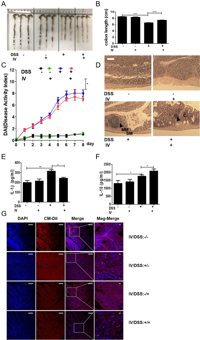
Figure 3.
Adoptive transfer of peritoneal macrophages from T. spiralis-infected mice inhibited DSS-induced colitis. After sacrificing the mice, colon length was measured (A,B). The percentage weight loss, changes in stool states including consistency, and blood presence in stools of mice were examined daily during the experimental period and represented as DAI. The DAI standard is shown in Table 2. The DAI results are shown in (C). The large intestines were isolated from mice, rolled up, fixed with 4% paraformaldehyde, stained with H&E, and examined for histopathological changes under a microscope (D). Arrows indicate sub-mucosal thickening and immune cell infiltration, characteristic of colitis. The immune cells of MLN were incubated for 72 h with anti-CD3 antibody (0.5 μg/mL) for T-cell stimulation. After incubation, cytokine production in the supernatant was determined by ELISA (E, F). Large intestine sections from mice receiving peritoneal macrophages (CM-Dil, red) were immunofluorescently stained for cell nuclei (DAPI, blue). Representative pictures are shown. White bar: 100 μm (G) Statistical analysis was performed with one-way ANOVA ((*p < 0.05, ***p < 0.001; n = 3 to 5 mice/group; these results are representative of three independent experiments).

