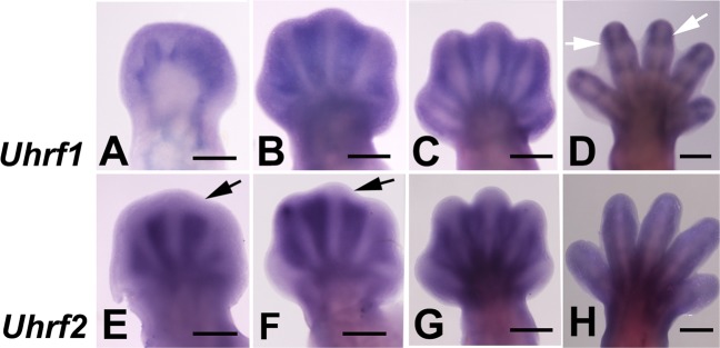
Fig. 2. In situ hybridizations showing the expression of Uhrf1 (a–d) and Uhrf2 (e–h) in the mouse autopod during interdigit remodeling.
a–d show the expression of Uhrf1 at pc days 12 (a), 13 (b), 13.5 (c), and 14 (d). Note the presence of joint domains by pc 14 (arrows in d). e–h show the expression of Uhrf2 at pc days 12.5 (e),13 (f), 13.5 (g), and 14 (h). Note the fading of the interdigital domains in the distal subectodermal region (arrows in e and f). Bars = 500 µm

