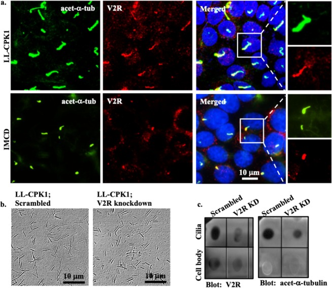
Figure 3.
V2R is localized to primary cilia. (a) Confocal images of renal epithelial from pig (LL-CPK1) and dog (IMCD) show localization of V2R (red) at the base of cilia. Acetylated-α-tubulin (acet-α-tub) used as ciliary marker is shown in green and nucleus in blue. Maximum intensity projection images from accumulated z-stack for LL-CPK1 (scrambled and V2R-knockdown), IMCD and endothelial cells are shown in Supplementary Figs S3 and S4. (b) Phase contrast images represent isolated cilia from scrambled control and V2R-knockdown LL-CPK1 cells. (c) Dot blot indicates the presence of V2R in both isolated-cilia and cell-body extracts. Isolated cilia extracts are confirmed by the presence of acetylated-α-tubulin.

