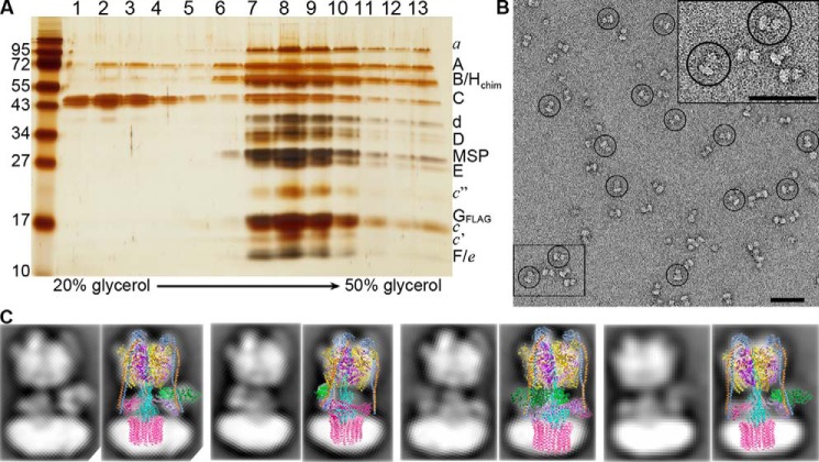
Figure 4.
Structural and functional characterization of the V1HchimVoND complex. A, reconstituted V1HchimVoND was subjected to glycerol gradient centrifugation, and the gradient fractions were analyzed by silver-stained SDS-PAGE. B, negative stain EM of V1HchimVoND showing homogeneous and monodisperse dumbbell-shaped molecules. Inset in the top right shows 2× zoomed area highlighted in the bottom left. C, a data set of ∼5800 particle projections was subjected to reference-free alignment and classification, and selected class averages were overlaid with projections of the cryoEM model of yeast V1Vo (Protein Data Bank code 3J9U). Bars in B, 50 nm.

