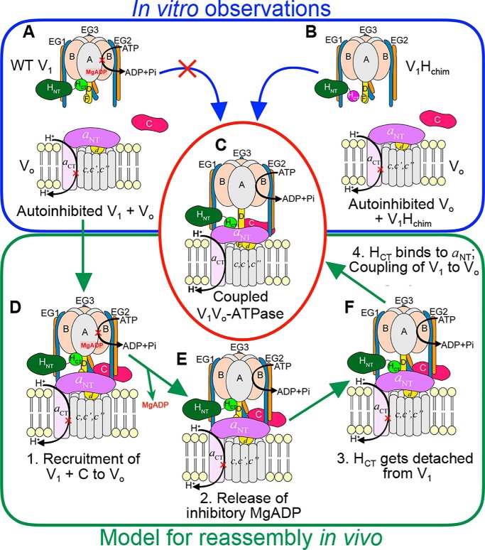
Figure 5.
Model for reassembly of autoinhibited V1 and Vo. A–C, our in vitro experiments have shown that although WT H containing V1 does not readily bind VoND (A), V1Hchim spontaneously associates with VoND (B) to form a structurally and functionally coupled V-ATPase, albeit at a slow rate. C, in vivo, however, V1 exists in the autoinhibited conformation (A), and the rate of assembly with Vo is significantly faster (within 5 min). D–F, for in vivo (re)assembly, we propose that the following steps occur: step 1, recruitment of V1 and subunit C to the vacuolar membrane (D); step 2, release of inhibitory MgADP (E); step 3, detachment of HCT from its inhibitory position on V1; and step 4, HCT binding to aNT (F). For further details, see text.

