Figure 1.
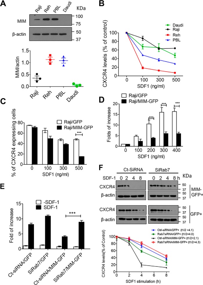
RAB7 is required for MIM-mediated CXCR4 internalization. A, immunoblot analysis of endogenous MIM protein in Raji, Reh, Daudi, and PBL cells. The expression levels were quantified based on the ratio of MIM to β-actin. B, malignant B cells were stimulated with SDF-1 at the indicated concentrations for 30 min. Surface expression of CXCR4 was analyzed by flow cytometry and normalized to that of control cells without SDF-1 treatment. C, Raji cells were transiently transfected with GFP or MIM-GFP and stimulated for 30 min with SDF-1 at different concentrations. The percentage of cells expressing surface CXCR4 was determined by flow cytometry. D, Raji cells expressing MIM-GFP or GFP were plated on Transwell plates in which the lower chamber was filled with medium containing SDF-1 at the indicated concentrations. After 24 h, cells were fixed and stained with 0.1% crystal violet. The number of cells that migrated to the lower chamber was compared with that of cells without SDF-1 treatment. E, HeLa cells expressing GFP or MIM-GFP were treated with siRAB7 or Ct-siRNA for 48 h. The treated cells were then subjected to a Transwell assay for their chemotactic response to 500 ng/ml SDF-1. F, HeLa cells expressing either MIM-GFP or GFP were treated with siRAB7 or Ct-SiRNA. The treated cells were further incubated with 500 μg/ml cycloheximide for 30 min prior to exposure to 150 ng/ml SDF-1 for the indicated times. The total amounts of CXCR4 protein in treated cells at different times were estimated by immunoblot and used to calculate t½ using Prism software. All data represent mean ± S.E.M. (n = 3). ***, p < 0.001.

